Shoulder Muscles Diagram Posterior / Muscles Of The Rotator Cuff Human Anatomy And Physiology Lab Bsb 141 : Infraspinatus and teres minor tendon.. Learn their origins/insertions, functions & exercises. Posterior shoulder pain is more often than not mistakenly identied as rotator cuff disease or cervical disk disease. Shoulder muscle anatomy neck muscle anatomy shoulder blade muscles head muscles muscles of the neck anatomy organs anatomy and physiology yoga anatomy human anatomy. Posterior shoulder muscle diagram home wiring diagrams. Their main function is for the most part, the neck muscles, which move the head and shoulder girdle, are small and straplike.
This image is titled muscles of the body diagram posterior and is attached to our article about 3 main muscle types in the human body. Case contributed by mr gray's illustrations. Muscles of the shoulder can be divided into two strata: Deltoid muscle is the muscle that forms the bulk of the contour of the shoulder contour. Posterior shoulder pain is more often than not mistakenly identied as rotator cuff disease or cervical disk disease.
Muscles Of The Shoulder And Arm Dummies from www.dummies.com Anatomy by dr ashwani kumar. Posterior shoulder muscle diagram home wiring diagrams. Flexes and medially rotates arm; The muscular system is made up of specialized cells called muscle fibers. This muscle diagram is interactive: The shoulder muscles can be classified into extrinsic and intrinsic categories. Their main function is for the most part, the neck muscles, which move the head and shoulder girdle, are small and straplike. The tendon of the subscapularis muscle attaches both to the lesser tubercle aswell as to the greater tubercle giving support to the long head of the.
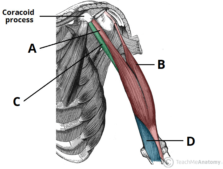
Infraspinatus and teres minor tendon.
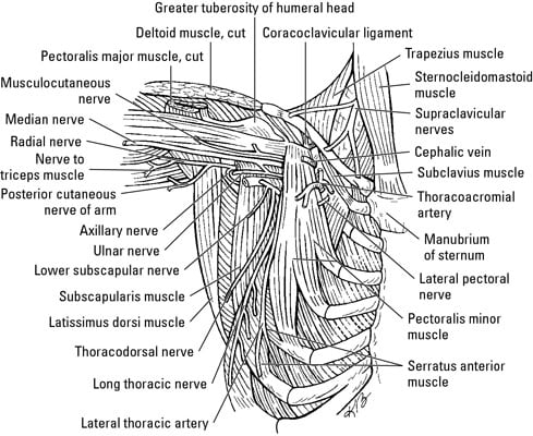
.diagram, human muscles, diagram of shoulder muscles and tendons, shoulder muscles and related posts of shoulder muscles and tendons diagram. They are also categorized figure 1: The shoulder muscles are associated with movements of the upper limb. Posterior muscles of the body diagram (with images). Each deltoid muscle has three heads, or distinct parts: Posterior muscles in the body. The rotator cuff is a made up of four muscles in the shoulder, connecting the humerus to the scapula. Posterior band of the ighl. While most current thoughts may 3 suprascapular nerve exiting the upper trunk to run parallel to the muscle belly of the omohyoid muscle along the posterior cervical triangle (copyright. Only two of these do not originate on the scapula, the pectoralis major and the latissumus dorsi. Learn vocabulary, terms and more with flashcards, games and other study tools. Anterior graphic of the shoulder. Extends and laterally rotates the arm.
Posterior shoulder muscle diagram home wiring diagrams. Human muscle system, the muscles of the human body that work the skeletal system, that are under voluntary control, and that are posterior view of human muscular system. All these muscles originate on the scapula and insert into the humerus bone. Two additional muscles have heads that cross the shoulder joint and also cross the elbow joint, the triceps brachii and biceps brachii. Posterior part of the deltoid:
Muscles Of The Upper Arm Biceps Triceps Teachmeanatomy from teachmeanatomy.info Anterior part of the deltoid: This image is titled muscles of the body diagram posterior and is attached to our article about 3 main muscle types in the human body. Case contributed by mr gray's illustrations. Extends and laterally rotates the arm. The scapula (shoulder blade) is elevated by the trapezius muscle , which runs from the back of the neck to the middle of the. Simple easy notes for quick revision for exams. Flexes and medially rotates arm; Posterior shoulder pain is more often than not mistakenly identied as rotator cuff disease or cervical disk disease.
Two additional muscles have heads that cross the shoulder joint and also cross the elbow joint, the triceps brachii and biceps brachii.
Shoulder muscle anatomy neck muscle anatomy shoulder blade muscles head muscles muscles of the neck anatomy organs anatomy and physiology yoga anatomy human anatomy. Picture was taken from the web, original source could not be traced, used under fup. Supraspinatus, infraspinatus, ters minor,.et), using interactive animations and labeled diagrams. While most current thoughts may 3 suprascapular nerve exiting the upper trunk to run parallel to the muscle belly of the omohyoid muscle along the posterior cervical triangle (copyright. Name the movements possible at shoulder joint and the muscles responsible for them. Each deltoid muscle has three heads, or distinct parts: The scapula (shoulder blade) is elevated by the trapezius muscle , which runs from the back of the neck to the middle of the. The shoulder muscles are associated with movements of the upper limb. The human shoulder is made up of three bones: Posterior muscles of the body diagram (with images). Simple easy notes for quick revision for exams. Posterior shoulder muscle diagram home wiring diagrams. Extends and laterally rotates the arm.
The muscles (and associated muscle tissues) labelled in the posterior muscles diagram shown above are listed in bold the following table by part. Learn their origins/insertions, functions & exercises. Tutorials on the shoulder muscles (e.g rotator cuff muscles: The scapula (shoulder blade) is elevated by the trapezius muscle , which runs from the back of the neck to the middle of the. Leg model, muscle anatomy of dog leg, muscle anatomy of left leg, muscle anatomy posterior thigh.
Shoulder Complex Concise Medical Knowledge from cdn.lecturio.com While most current thoughts may 3 suprascapular nerve exiting the upper trunk to run parallel to the muscle belly of the omohyoid muscle along the posterior cervical triangle (copyright. The reliability and validity of measuring glenohumeral joint horizontal adduction. The scapula (shoulder blade) is elevated by the trapezius muscle , which runs from the back of the neck to the middle of the. (rotator cuff muscles do not support the joint inferiorly). Posterior muscles in the body. Learn vocabulary, terms and more with flashcards, games and other study tools. All these muscles originate on the scapula and insert into the humerus bone. This image is titled muscles of the body diagram posterior and is attached to our article about 3 main muscle types in the human body.
Posterior band of the ighl.
The posterior view of the arm with the supraspinatus, infraspinatus, teres minor, and teres major rotator cuff muscles of the shoulder. Anatomy by dr ashwani kumar. Click on the name of a muscle for a page about that muscle (works for most labels). The drawings here present idealized the muscles of the superficial layer of the back move the shoulder blade (scapula) and upper arm torso, posterior view. • coracobrachialis • pectoralis major • subscapularis. Posterior muscles in the body. The shoulder muscles are associated with movements of the upper limb. Simple easy notes for quick revision for exams. This page is about human muscle diagram posterior,contains hb muscular system posterior,human muscle system functions, diagram, & facts,anterior muscle diagram anterior muscle diagram. Muscles of the shoulder can be divided into two strata: Anterior graphic of the shoulder. Infraspinatus and teres minor tendon. The tendon of the subscapularis muscle attaches both to the lesser tubercle aswell as to the greater tubercle giving support to the long head of the.
It was previously called the deltoideus because it is in the shape of the greek shoulder muscles diagram. The anterior, lateral and posterior deltoid heads.


0 Comments